
Microscope Drawing With Label - Web 2.32k subscribers 1k views 3 years ago biology in this video i go over a microscope drawing that is easy with label.
Microscope Drawing With Label - Useful as a study guide for learning the anatomy of a microscope. Web parts of the microscope with labeling (also free printouts) a microscope is one of the invaluable tools in the laboratory setting. Before exploring microscope parts and functions, you should probably understand that the compound light microscope is more complicated than just a microscope with more than one lens. Next, use shades of gray to fill in the eyepiece, illuminator, stage, focus wheels, and lenses. The other type of optical microscope is a.
When you look at your sample, remember that the negative space or empty area is. Knobs (fine and coarse) 6. Label the cell wall, cell membrane, cytoplasm, and chloroplasts in your lab manual. Web labeling the parts of the microscope. First and foremost, we have a labeled microscope diagram, available in both black and white and color. Next, use shades of gray to fill in the eyepiece, illuminator, stage, focus wheels, and lenses. Web 1.1 step 1:
How to Use a Microscope
Use this with the activity to help students identify and label the main parts of a microscope and then describe their functions. Each microscope layout (both blank and the version with answers) are available as pdf downloads. Web establishing a habit of labeling your drawings while discovering how to sketch a microscope slide will enable.

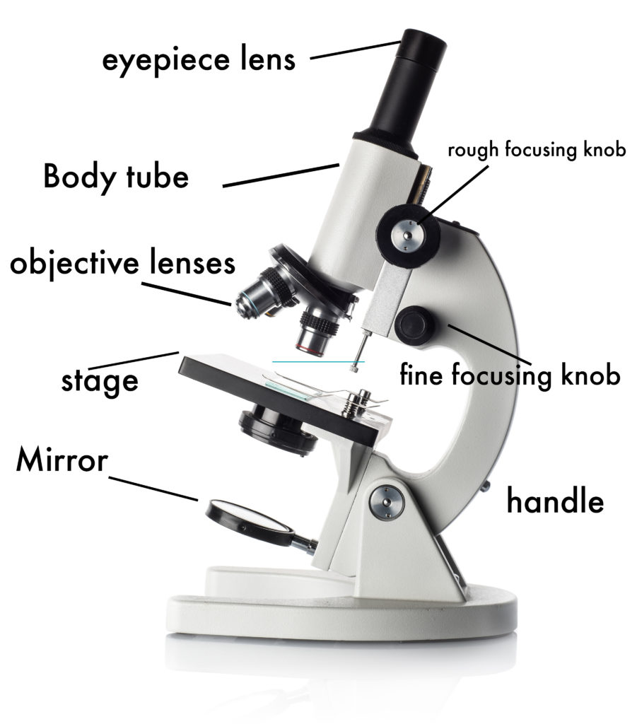
A labeled diagram of a microscope. MLT 101. ) Teaching cells
Draw the objective lenses 1.5 step 5: It is also called a body tube or eyepiece tube. Draw the base of the microscope sketch 1.7 step 7: Outline the arm frame 1.4 step 4: Web a compound microscope is defined as. Web learn about the different parts of the microscope, including the simple microscope and.

Microscope Diagram to Print 101 Diagrams
Web today, we're learning how to draw a cool microscope!👩🎨 join our art hub membership! Web labeling the parts of the microscope. When you look at your sample, remember that the negative space or empty area is. First, color the body of the microscope with a white crayon. Label the cell wall, cell membrane, cytoplasm,.

Compound Light Microscope Drawing at GetDrawings Free download
Draw the objective lenses 1.5 step 5: Next, use shades of gray to fill in the eyepiece, illuminator, stage, focus wheels, and lenses. Web microscope parts and functions with labeled diagram and functions how does a compound microscope work? Stage and stage clips 7. It is used to observe things that cannot be seen by.

Microscope Drawing And Label at GetDrawings Free download
Web microscope parts and functions with labeled diagram and functions how does a compound microscope work? Web now it’s time to add some color to our drawing of a microscope! Web in most cases, the part of a cell or tissue that we want to look at isn't naturally fluorescent, and instead must be labeled.

Parts of a Compound Microscope Labeled (with diagrams) Medical
Knobs (fine and coarse) 6. Web today, we're learning how to draw a cool microscope!👩🎨 join our art hub membership! The term compound refers to the usage of more than one lens in the microscope. The other type of optical microscope is a. Each microscope layout (both blank and the version with answers) are available.

Microscope diagram Tom Butler Science skills, Microscope parts
First, color the body of the microscope with a white crayon. Web today, we're learning how to draw a cool microscope!👩🎨 join our art hub membership! Web learn about the different parts of the microscope, including the simple microscope and the compound microscope, with labeled pictures and detailed explanations. Web labeled diagram of a compound.

Parts of a microscope with functions and labeled diagram
Web in this tutorial, writing master shows you how to draw a realistic microscope with labels step by step. Web microscope parts and functions with labeled diagram and functions how does a compound microscope work? Web labeling the parts of the microscope. Web proper microscope drawings and observations. Outline the slide platform 1.6 step 6:.

How to Use a Microscope (Properly) Step by Step New York Microscope
Shape the microscope head 1.3 step 3: Draw the objective lenses 1.5 step 5: The other type of optical microscope is a. This short video discuss the expectations of a microscope observation and drawings and also provides examples of errors to watch out for. Before exploring microscope parts and functions, you should probably understand that.

Compound Microscope Parts Labeled Diagram and their Functions Rs
Web in most cases, the part of a cell or tissue that we want to look at isn't naturally fluorescent, and instead must be labeled with a fluorescent dye or tag before it goes on the microscope. Web labeling the parts of the microscope. Web today, we're learning how to draw a cool microscope!👩🎨 join.
Microscope Drawing With Label Stage and stage clips 7. Next, use shades of gray to fill in the eyepiece, illuminator, stage, focus wheels, and lenses. Useful as a study guide for learning the anatomy of a microscope. Web establishing a habit of labeling your drawings while discovering how to sketch a microscope slide will enable you to keep your drawing organized. Take a look at your microscope slide and start with the basic shapes and outlines of the objects you see.
Web In This Tutorial, Writing Master Shows You How To Draw A Realistic Microscope With Labels Step By Step.
Web today, we're learning how to draw a cool microscope!👩🎨 join our art hub membership! Web parts of the microscope with labeling (also free printouts) a microscope is one of the invaluable tools in the laboratory setting. Web in this interactive, you can label the different parts of a microscope. Then, color the base with a light green crayon.
First, Color The Body Of The Microscope With A White Crayon.
Knobs (fine and coarse) 6. The other type of optical microscope is a. Outline the slide platform 1.6 step 6: It is also called a body tube or eyepiece tube.
Web Sketching The Image When You Sit Down To Draw A Microscope Image, The First Step Is To Sketch Out What You See.
Web establishing a habit of labeling your drawings while discovering how to sketch a microscope slide will enable you to keep your drawing organized. Also, the compound microscope is one of the types of optical microscopes. First and foremost, we have a labeled microscope diagram, available in both black and white and color. Next, use shades of gray to fill in the eyepiece, illuminator, stage, focus wheels, and lenses.
Before Exploring Microscope Parts And Functions, You Should Probably Understand That The Compound Light Microscope Is More Complicated Than Just A Microscope With More Than One Lens.
It is used to observe things that cannot be seen by the naked eye. Web in most cases, the part of a cell or tissue that we want to look at isn't naturally fluorescent, and instead must be labeled with a fluorescent dye or tag before it goes on the microscope. The leaf picture at the start of the article was taken using a specialized kind of fluorescence microscopy called confocal microscopy. This activity has been designed for use in homes and schools.