
Spinal Cord Drawing - Web the spinal cord begins at the base of the brain and extends into the pelvis.
Spinal Cord Drawing - It carries signals between the brain and the rest of the body. Web the spinal cord is a long, thin, tubular structure made up of nervous tissue that extends from the medulla oblongata in the brainstem to the lumbar region of the vertebral column (backbone) of vertebrate animals.the center of the spinal cord is hollow and contains a structure called central canal, which contains cerebrospinal fluid.the spinal cord is also. Ventral horn, dorsal horn, white matter, gray matter, meninges, central canal, dorsal root ganglion, dorsal root of the spinal nerve, and the ventral root of the spinal nerve. Cervical, thoracic, lumbar and sacral, two of these are marked by an upper (cervical) and a lower (lumbar) enlargement. A vector illustration of a blueprint of the human spine.
A vector illustration of a blueprint of the human spine. Human joints and body parts bones sketch icons. This kind of long tube that runs down the spine. Diagram of the spinal cord is illustrated in detail with neat and clear labelling. A number of approaches exist to improve learning and retention of neuroanatomy and clinical localization principles. Spine isolated on a white backgrounds. Representation in 3/4 front view of the stucture of the spinal cord, and rachidian nerves.

Spinal Cord Anatomy Nurse Info
These nerve signals help you feel sensations and move your muscles. Web the module promotes learning and mastery of spinal cord anatomy and lesion localization. Diagram of the spinal cord is illustrated in detail with neat and clear labelling. Spine isolated on a white backgrounds. A vector illustration of a blueprint of the human spine..

Spinal cord diagram
Web the spinal cord is a long bundle of nerves and cells that extends from the lower portion of the brain to the lower back. Learn more about the spinal cord with our learning material. Web the spinal cord runs through a hollow case from the skull enclosed within the vertebral column. This kind of.

The Spinal Cord Neurologic Clinics
Diagram of the spinal cord is illustrated in detail with neat and clear labelling. Spinal cord drawing stock photos are available in a variety of sizes and formats to fit your needs. These nerve signals help you feel sensations and move your muscles. Vector sketch icons of human body bones and joints. A vector illustration.

Spinal Cord Anatomy, Structure, Function, & Diagram
Human joints and body parts bones sketch icons. Your spinal cord helps carry electrical nerve signals throughout your body. Web according to its rostrocaudal location the spinal cord can be divided into four parts: It carries signals between the brain and the rest of the body. Web each segment of the spinal cord provides several.

Human Spinal Cord Drawing Sketch Coloring Page
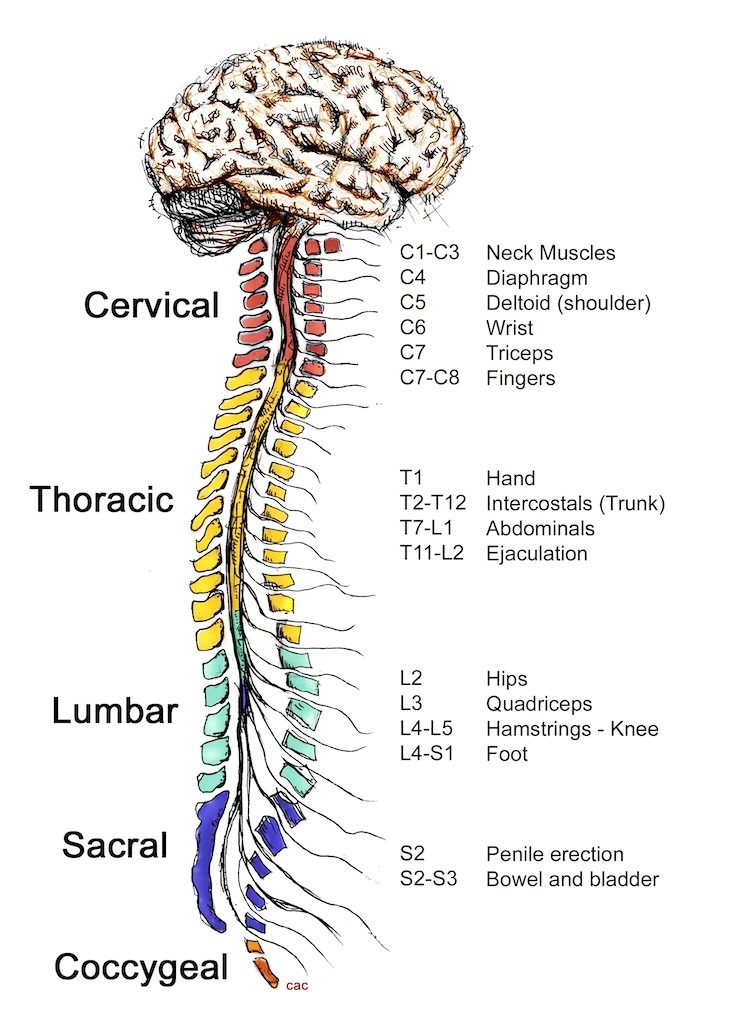
There are 8 pairs of cervical, 12 thoracic, 5 lumbar, 5 sacral, and 1 coccygeal pair of spinal nerves (a total of 31 pairs). Web the module promotes learning and mastery of spinal cord anatomy and lesion localization. Here i upload carefully crafted videos to meet the problems in drawing.all the videos are. Web vector.

The spinal cord Anatomy of the spinal cord Physiology of the spinal
On each half of the spinal cord, a ventrolateral and dorsolateral sulcus is appreciated at the sites from which the ventral and dorsal nerve roots leave and enter the spinal cord. A number of approaches exist to improve learning and retention of neuroanatomy and clinical localization principles. Hand drawn spine isolated on a white backgrounds..

11.1A Overview of the Spinal Cord Medicine LibreTexts
Web the module promotes learning and mastery of spinal cord anatomy and lesion localization. Here i upload carefully crafted videos to meet the problems in drawing.all the videos are. Only show results related to: Web the spinal cord is a long bundle of nerves and cells that extends from the lower portion of the brain.

Anatomy of the Spinal Cord Praxis Spinal Cord Institute
Web use this line drawing to refresh your understanding of the gross anatomy of the spinal cord, paying particular attention to the following structures in this figure: Hand drawn spine isolated on a white backgrounds. Web each segment of the spinal cord provides several pairs of spinal nerves, which exit from vertebral canal through the.
How Does The Spinal Cord Work Reeve Foundation
Welcome to my just made easy official youtube channel. Cervical enlargement, lumbosacral enlargement, medullary cone, spinal part of filum terminale, cauda equina, spinal nerves. Web the module promotes learning and mastery of spinal cord anatomy and lesion localization. Let us learn how to draw a human. Here i upload carefully crafted videos to meet the.

Anatomy and Health Charts Free Printable PDF Files Human body anatomy
It contains tissues, fluids and nerve cells. Web the module promotes learning and mastery of spinal cord anatomy and lesion localization. And there's a number of structures coming out of the spinal cord that i'll talk about next. Web according to its rostrocaudal location the spinal cord can be divided into four parts: These nerve.
Spinal Cord Drawing Representation in 3/4 front view of the stucture of the spinal cord, and rachidian nerves. Web the spinal cord can be equally divided along the midline dorsoventral axis by drawing a line through the depression known as the dorsal and ventral median sulci. Web here's a drawing of the spinal cord. Here i upload carefully crafted videos to meet the problems in drawing.all the videos are. This kind of long tube that runs down the spine.
On Each Half Of The Spinal Cord, A Ventrolateral And Dorsolateral Sulcus Is Appreciated At The Sites From Which The Ventral And Dorsal Nerve Roots Leave And Enter The Spinal Cord.
A number of approaches exist to improve learning and retention of neuroanatomy and clinical localization principles. Web the module promotes learning and mastery of spinal cord anatomy and lesion localization. Diagram of the spinal cord is illustrated in detail with neat and clear labelling. Human joints and body parts bones sketch icons.
Representation In 3/4 Front View Of The Stucture Of The Spinal Cord, And Rachidian Nerves.
Web the spinal cord is a long bundle of nerves and cells that extends from the lower portion of the brain to the lower back. Web vector human spine blueprint. Web the spinal cord is a long, thin, tubular structure made up of nervous tissue that extends from the medulla oblongata in the brainstem to the lumbar region of the vertebral column (backbone) of vertebrate animals.the center of the spinal cord is hollow and contains a structure called central canal, which contains cerebrospinal fluid.the spinal cord is also. Welcome to my just made easy official youtube channel.
And There's A Number Of Structures Coming Out Of The Spinal Cord That I'll Talk About Next.
Web the module promotes learning and mastery of spinal cord anatomy and lesion localization. Learn more about the spinal cord with our learning material. Web spinal cord, drawing the spinal cord. A vector illustration of a blueprint of the human spine.
A Number Of Approaches Exist To Improve Learning And Retention Of Neuroanatomy And Clinical Localization Principles.
Web your spinal cord is the long, cylindrical structure that connects your brain and lower back. Web use this line drawing to refresh your understanding of the gross anatomy of the spinal cord, paying particular attention to the following structures in this figure: A bony column of vertebrae surrounds and protects your spinal cord. Ventral horn, dorsal horn, white matter, gray matter, meninges, central canal, dorsal root ganglion, dorsal root of the spinal nerve, and the ventral root of the spinal nerve.